
Figure 1.
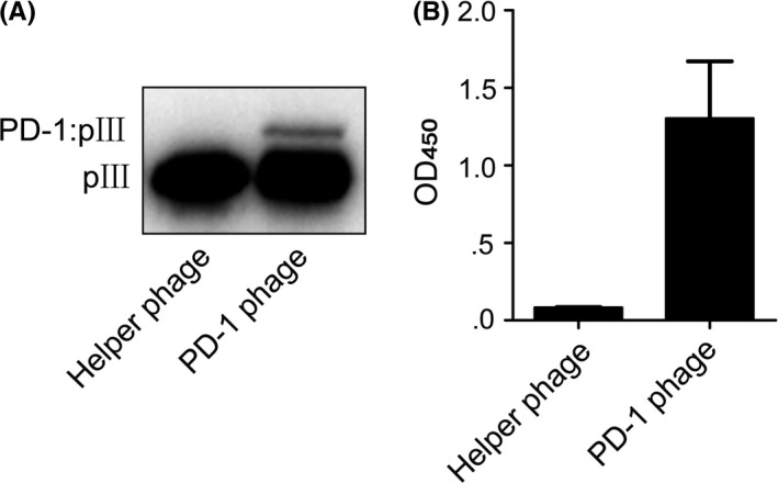
Display of hPD‐1 extracellular region on M13 phage. A, Western blot of PD‐1 phage. B, The binding of PD‐1 phage to hPD‐L1 detected by ELISA. Helper phage was run as a negative control. Error bars indicate SD (n = 3)
Official websites use .gov
A
.gov website belongs to an official
government organization in the United States.
Secure .gov websites use HTTPS
A lock (
) or https:// means you've safely
connected to the .gov website. Share sensitive
information only on official, secure websites.
Display of hPD‐1 extracellular region on M13 phage. A, Western blot of PD‐1 phage. B, The binding of PD‐1 phage to hPD‐L1 detected by ELISA. Helper phage was run as a negative control. Error bars indicate SD (n = 3)