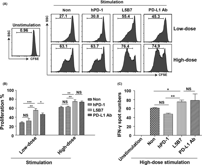
Figure 6.

The enhancement of proliferation and IFN‐γ release of activated PBMC. A, The flow cytometry data of proliferation of activated PBMC with anti‐CD3 Ab (aCD3) and anti‐CD28 Ab (aCD28) in the presence or absence of soluble hPD‐1, L5B7 and anti‐PD‐L1 Ab (5 μg/mL). PBMCwere stimulated with low‐dose antibodies of 15 ng/mL aCD3 and 7.5 ng/mL aCD28 or high‐dose antibodies of 30 ng/mL aCD3 and 15 ng/mL aCD28. B, The statistical data of proliferating cells in (A). Error bars indicated SD (n = 3). C, ELISpot assays showing IFN‐γ release of PBMC activated with 30 ng/mL aCD3 and 15 ng/mL aCD28 in the presence or absence of soluble hPD‐1, L5B7 and anti‐PD‐L1 Ab (5 μg/mL). Error bars indicated SD (n = 3). Unpaired Student's t test, NS, P > .05; *, P < .05; **, P < .01; ***, P < .001
