Figure 5.

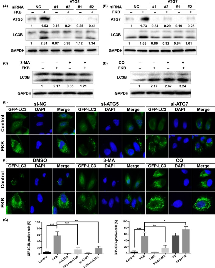
Flavokawain (FKB) induces autophagic flux in thyroid cancer cells. (A,B) ARO, WRO, and TPC‐1 cells were transfected with ATG5 or ATG7 siRNAs for 24 h, and treated with 6 μg/mL FKB or DMSO for another 24 h. The samples were subjected to Western blot analysis. Levels of protein expression were analyzed by Western blot using antibodies against ATG5, ATG7, LC3B, and GAPDH. (C,D) ARO, WRO, and TPC‐1 cells were pretreated with 3‐methyladenine (3‐MA) (10 mmol/L) or chloroquine (CQ) (3 μmol/L) for 1 h, and co‐incubated with 6 μg/mL FKB or DMSO for another 24 h. Levels of protein expression were analyzed by Western blot using antibodies against LC3B and GAPDH. (E) WRO cells stably transfected with GFP‐LC3B were further transfected with ATG5 or ATG7 siRNAs, then treated with 6 μg/mL FKB or DMSO for another 24 h. Cells were then subjected to a fluorescence microscopy. (F) WRO cells stably transfected with GFP‐LC3B were cotreated with autophagy inhibitors and FKB as mentioned above, then examined by a fluorescence microscopy. (G) Percentages of cells with more than four GFP‐LC3B dots were quantified. All data are expressed as the mean ± SD. *P < 0.05; **P < 0.01; ***P < 0.001
