Figure 1.

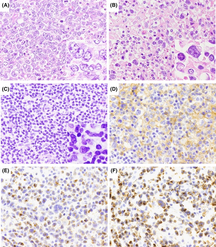
Light microscopy images of nodal Epstein‐Barr virus ‐negative cytotoxic molecule(CM)‐positive peripheral T‐cell lymphoma samples. Hematoxylin and eosin staining was performed to examine nuclear morphology, revealing centroblastoid morphology (A), pleomorphic morphology (B) and mixed morphology (C). Other samples were immunostained for CD4 (D), TIA‐1 (E), and granzyme B (F). Original magnification: 400×. Inset: enlarged view of tumor cells
