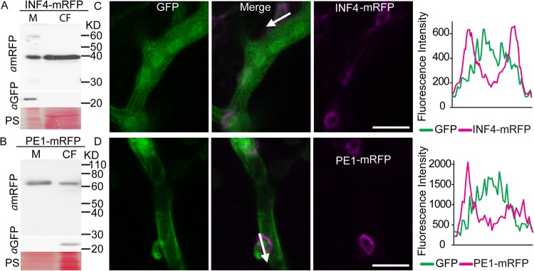
FIG 1 .
Phytophthora infestans elicitor-like protein INF4 and cell wall-degrading enzyme pectinesterase PE1 are secreted from mycelium into the culture filtrate (CF) in vitro and at haustoria in planta. (A and B) Ham34 promoter-driven constitutive expression of the INF4 and PE1 fusion proteins tagged with monomeric red fluorescent protein (mRFP) in the mycelium (M) and secretion into the CF of in vitro-grown P. infestans were confirmed using an mRFP antibody. Enhanced green fluorescent protein (EGFP; detected with aGFP) was used as a marker of cytoplasmic protein, and the results showed that the CF preparation was not detectably contaminated with cellular protein. Size markers are indicated in kilodaltons, and protein loading is indicated by Ponceau staining (PS). (C and D) Confocal projections of P. infestans transformants expressing GFP in the hyphal cytoplasm and INF4-mRFP (C) or PE1-mRFP (D). Secretion of INF4 and PE fusions was observed particularly at haustoria in planta. The white arrows show the lines used to generate the fluorescence intensity profiles indicated in the graphs to the right of the images. The x-axis data in the graphs represent the distances (in micrometers) from one end of each white arrow in the images to the other end. Scale bars represent 10 µm.

