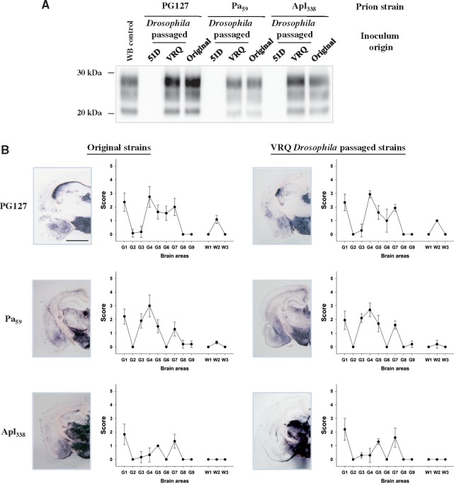
Figure 4.
Authentic prion replication in PrP transgenic Drosophila. Actin × VRQ(GPI) PrP transgenic (VRQ) Drosophila were exposed to the PG127 and Apl338, prion strains, and Elav × ARQ(GPI) PrP transgenic (ARQ) Drosophila were exposed to the Pa59 prion strain, at the larval stage. Actin × 51D and Elav × 51D (both referred to as 51D in appropriate graph) Drosophila were used as control flies where appropriate. Head homogenates were prepared from adult flies aged 30 days and inoculated into tg338 mice. Inoculated mice were euthanized when they showed clinical signs of prion infection or after 250 days for those that did not develop clinical disease. Mice were considered positive for prion disease when PK-resistant PrP27–30 was detected in brain tissue by western blot. Mouse brains were analysed for the presence of PK-resistant PrP and subjected to neuropathological assessment by PET blot and lesion profile analysis. (A) Western blot detection of PK-resistant PrP27–30 in the brains of tg338 mice during the isolation of PG127, Pa59 or Apl338, or prion strains (original inoculum), or these prion strains after passage in Drosophila (Drosophila passaged). Molecular mass markers in kDa are shown on the left. (B) PET blot and lesion profile analysis of tg338 mouse brains after exposure to the original mouse-derived PG127, Pa59 or Apl338, or prion strains (original inoculum), or these prion strains after passage in Drosophila (Drosophila passaged). Scale bar = 150 µm.

