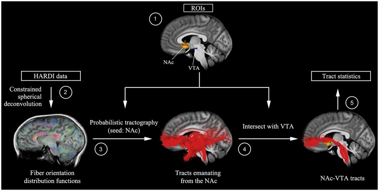
Figure 1.
Overall HARDI mesolimbic reward pathway analysis pipeline. (1) NAc region of interest (ROI) was demarcated using FreeSurfer-based segmentations of each individual’s T1-weighted anatomical image, and then warped to diffusion space using parameters from a nonlinear co-registration between T1 and fractional anisotropy images implemented in the advanced normalization tools software package (Avants et al., 2011). A VTA region of interest was determined using midbrain activation peaks from a reward (gambling) task performed by 448 participants as part of the Human Connectome Project (Van Essen et al., 2012)—this approach allowed us to reliably identify VTA voxels that are most sensitive to reward. VTA regions of interest were transformed to individual subject diffusion space using a 2D template-based approach to maximize anatomical precision (see Supplementary material for details). (2) Fibre orientation distribution functions were computed by constrained spherical deconvolution with a maximum harmonic degree of 10, using MRtrix3 (Tournier et al., 2012). (3) Probabilistic tractography was then performed by seeding 10 000 points in each voxel of the NAc and generating streamlines using the iFOD2 algorithm implemented in MRtrix3 (Tournier et al., 2010). (4) Tracts emanating from the NAc were intersected with ipsilateral VTA region of interest to identify NAc-VTA tracts. (5) Fibre density—the target-volume (VTA) normalized fraction of streamlines emanating from the NAc and intersecting ipsilateral VTA—was then computed for each child.

