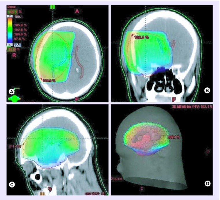
Figure 2. . 3D dose distribution.
Dose-wash in (A) axial, (B) coronal and (C) sagittal sections with (D) 3D dose reconstruction on a planning CT scan. Note that coverage of the planning target volume (in red) by 95% isodose (in green) results in high-dose irradiation of the ipsilateral SVZ (in blue) while sparing most of the contralateral SVZ (in brown).

