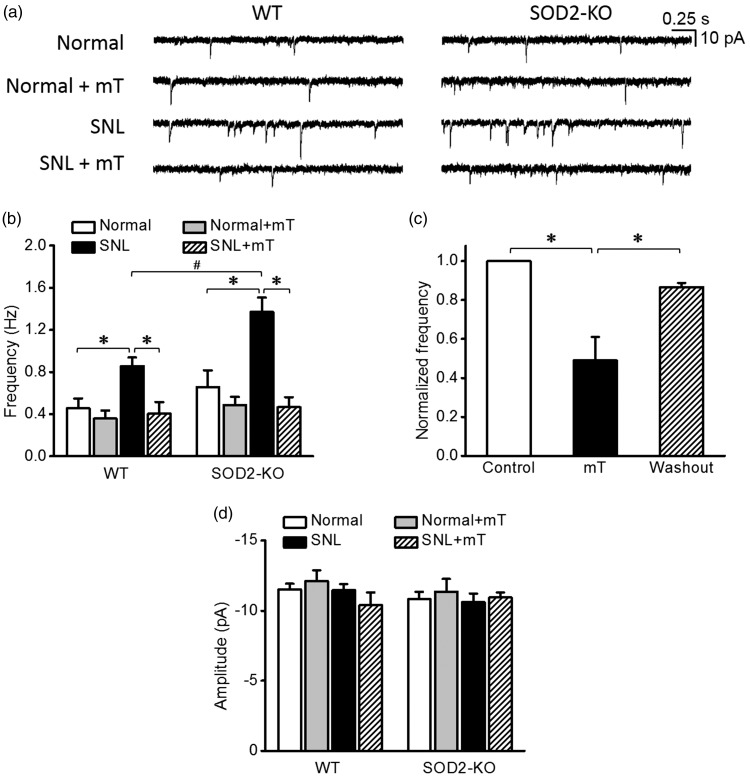
Figure 2.
Miniature excitatory postsynaptic currents (mEPSCs) in spinal dorsal horn neurons of normal and neuropathic SOD2-KO mice and their WT littermates. (a) Representative traces of mEPSCs in normal and SNL groups before and during the perfusion of mitochondria-targeting superoxide scavenger, mitoTEMPO (mT, 300 μM). (b) The summary graphs showing the mean frequency of the mEPSCs in each group. SNL significantly increased mEPSC frequency in both WT and SOD2-KO mice. Notably, this SNL-increased mEPSC frequency was greater in SOD2-KO mice than in their WT counterparts. These increased frequencies returned to the normal level by mT in a reversible manner (c, n = 8 from three mice). No difference in mEPSC amplitude was detected between groups (d). *p < 0.05 between control vs. mT, mT vs. washout, normal vs. SNL, and SNL vs. SNL+mT; #p < 0.05 between SNL WT mice vs. SNL SOD2-KO mice by two-way analysis of variance followed by Holm–Sidak multiple comparison tests. WT: wild-type; SNL: spinal nerve ligation.

