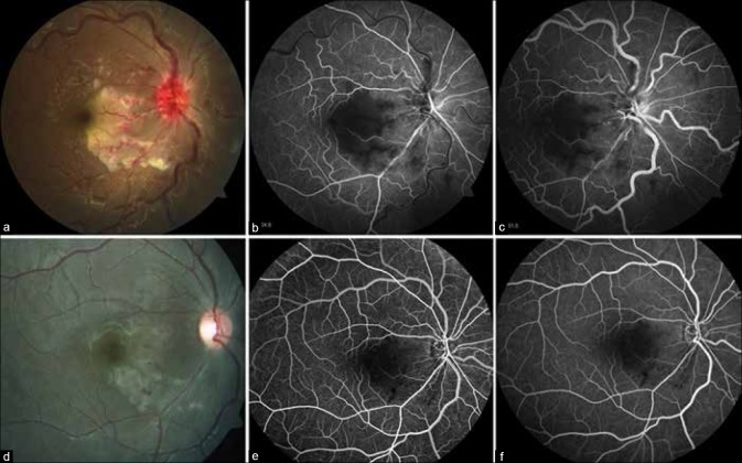
Figure 2.
(a) Color fundus photograph of right eye shows a circumscribed area of retinal whitening abutting fovea with hyperemic and edematous optic disc, marked venous tortuosity along with retinal hemorrhages, and cotton wool spots. Fluorescein angiogram of the right eye showing widespread arteriolar occlusion and capillary non-perfusion persisting in (b) early and (c) late phases. (d) At 3 weeks, disc edema resolved, hemorrhages and retinal whitening reduced along with narrowing of the arterioles, with improved perfusion of the arterioles and capillary plexus (e and f)

