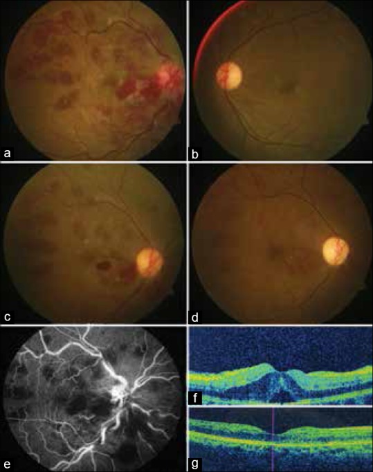
Figure 1.

(a and b) Posterior pole fundus photograph of right and left eye at initial presentation, (c and d) images of the right eye at subsequent follow-up at 2 monthly intervals after change of diet plan with regard to celiac disease and subsequent clearing of the retinal hemorrhages; (e) the mid arteriovenous phase of fundus fluorescein angiography shows blocked fluorescence due to hemorrhages; (f) the presence of cystoid macular edema at fovea, and (g) the resolution of cystoid macular edema postintravitreal injection of bevacizumab
