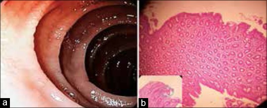
Figure 4.

(a) Areas of duodenal erosions with scalloping of duodenal folds and loss of villi, (b) histopathology of duodenal biopsy showing signs of atrophic villi, decreased villous/crypt ratio (<1.1), crypt hyperplasia, increased intraepithelial lymphocytes, and lamina propria showing moderate infiltration with plasma cells, (H and E, ×200)
