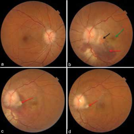
Figure 1.

(a and b) Fundus photograph of both eye at presentation, showing hyperemic optic discs with blurred margins and presence of a 4 disc diameter area of subretinal hemorrhage (red arrow) inferotemporal to the optic disc in the left eye along with subretinal fluid involving the fovea (green arrow) and a 1/3 disc diameter choroidal neovascular membrane complex just temporal to the optic disc (black arrow), (c) 3 weeks after intravitreal ranibizumab decrease in subretinal hemorrhage with healing of choroidal neovascular membrane (red arrow), (d) at 3 months there is complete resolution of hemorrhage and fluid with a scarred choroidal neovascular membrane (red arrow)
