
Figure 4.
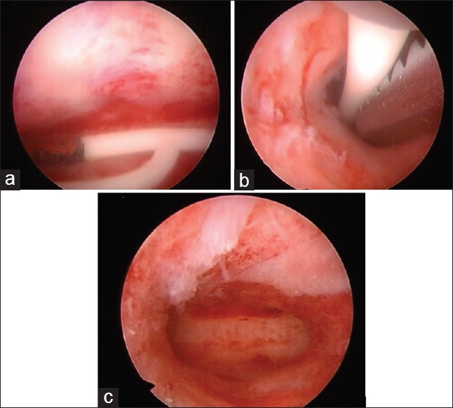
Hysteroscopic pictures. Retained intrauterine device for 16 years in a 51-year-old G3P3 (3003). Note the rusted areas on the short arm (a). Removal done using a semirigid Fr. 3 grasping forceps (b). Endometrium after removal of the intrauterine device. Note the indentation due to the device at the fundal area (c)