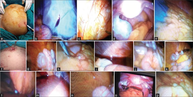
Figure 4.
Palmar point entry (a), trocar puncture of mass for decompression (b), peritoneal implants not detected by imaging (c and n), ovarian mass with Douglas pouch evaluation and free intestine (d), free omentum (e), Everting the umbilicus for entry in pelvic masses (f), ovarian mass (g), omental deposits not detected by imaging (h), normal diaphragm with hemorrhagic ascites (i), normal intestine and mesentery (j and k) Falciform deposits not detected on imaging (l), normal liver (m), oophorectomy with other side biopsies (o), omental deposits not detected by imaging (p)

