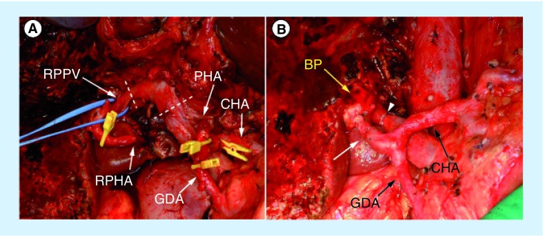
Figure 4. Intraoperative photographs during the left hepatic trisectionectomy with caudate lobectomy.
The portal vein is circularly transected at the dotted lines (A) and was reconstructed in direct end-to-end anastomosis (B) with an arrowhead. The hepatic arterial end-to-end anastomosis between the right posterior hepatic artery and proper hepatic arteries is indicated (B) with an arrow. The dashed lines present the resection line of the portal vein.
BP: Bile duct stump of the right posterior sectional duct; CHA: Common hepatic artery; GDA: Gastroduodenal artery; PHA: Proper hepatic artery; RPHA: Posterior branch of the right hepatic artery; RPPV: Right posterior portal vein.

