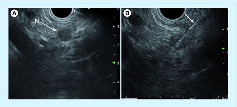
Figure 6. Endoscopic ultrasonography and fine-needle aspiration cytology under endoscopic ultrasonography guidance.
(A) Endoscopic ultrasonography reveals a hypoechoic nodule at the lesser curvature of the stomach. The endoscopic ultrasonography-fine-needle aspiration cytology for a swollen lymph node showed adenocarcinoma. (B) The arrow indicates endoscopic ultrasonography-fine-needle aspiration needle.
LN: Lymph node.

