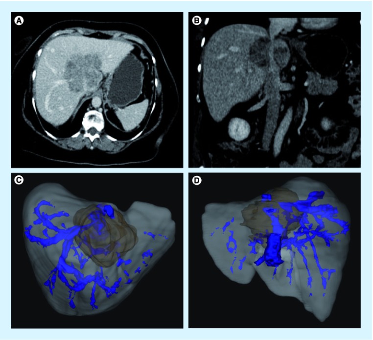
Figure 4. Use of 3D imaging to plan an inferior vena cava and hepatic vein resection of an intrahepetic cholangiocarcinoma.
(A & B) The location of the tumor on standard triphasic CT scan imaging. 3D imaging from (C) a crainocaudal view and (D) from the posterior view. High-quality imaging is essential for complex vascular reconstructions.

