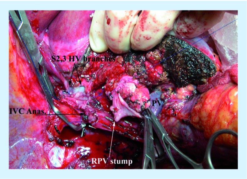
Figure 6. Right trisectionectomy, using cold perfusion to facilitate resection of the hepatic vein confluence and reanastomosis of the inferior vena cava and the anastomosis of the segment 2 and 3 branches of the left hepatic vein to the inferior vena cava.
The University of Wisconsin solution was instilled through the stump of the right portal vein with outflow through the hepatic veins prior to reanastamosis. At the time this picture was taken, the back wall of the hepatic vein and inferior vena cava anastomosis was completed, after the segment 2 and 3 branches were plastied together to form a common orifice. Prior to completion of the anastomosis, the liver was flushed with lactated ringers containing 25% albumin to wash out the University of Wisconsin solution.
Anas: Anastomosis; HV: Hepatic vein; IVC: Inferior vena cava; PV: Portal vein; RPV: Right hepatic vein.

