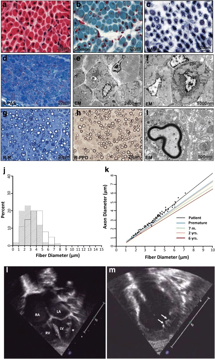
Fig. 1.
a-i Muscle biopsy performed at age of eight weeks shows a myopathy with increased variation in muscle diameter and internalized nuclei at H&E (a) and Gomori trichrome (b) stained cryosections consistent with a centronuclear myopathy. In c NADH staining the muscle fibers show disturbance of the internal myofiber architecture with central dark staining and pale surrounding halo. Many fibers show increased glycogen in PAS-stained semithin sections (d) and at ultrastructural analyses with disturbance of the myofiber architecture (e, f). In the sural nerve biopsy, the number of myelinated fibers appears slightly decreased (g) and the axons have mostly thin myeline sheats (h, i). j-k Morphometric analyses of the sural nerve biopsy at age eight weeks. j The histogram shows the frequency of distribution of axonal (hatched) and fiber diameters. The diameter distribution is unimodal with a diameter between 1 and 8 μm and shows an increased frequency of small fibers and axons for this age. k Analyzing the correlation of fiber to axonal diameter the patient has thinner myelin sheaths (slope = 0.87) compared to normal controls in literature [3, 7] (slope = 0.77) (m = month, yrs. =years). l-m Echocardiography of the patient at age 10 weeks. Four-chamber view (l) and isolated view on the left ventricle (m) demonstrating enlarged atria as suggestive of restrictive cardiac dysfunction, and abnormal trabeculation and intra-trabecular recesses (arrows and asterisk) as characteristic of left-ventricular non-compaction cardiomyopathy

