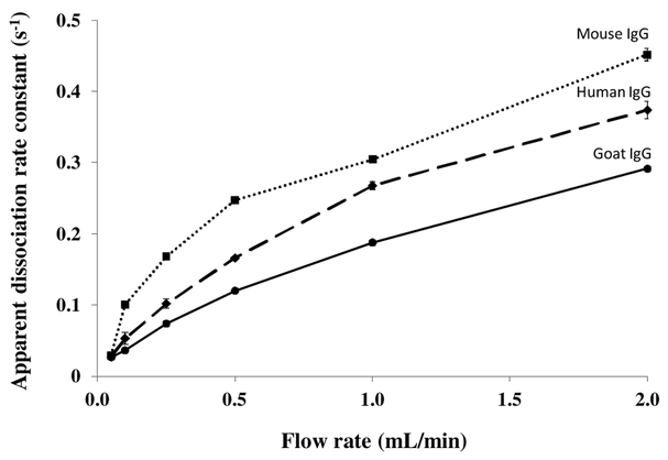
Figure 5.
Apparent dissociation rate constants measured by the peak decay method for goat (●), human (♦) and mouse (■) IgG as a function of elution flow rate. These results were obtained using a 5 mm × 2.1 mm I.D. protein G microcolumn and a pH 2.5 elution buffer that was passed through the microcolumn at 0.05–2.0 mL/min. The results obtained for rabbit IgG were given in Figure 4. The error bars represent ± 1 standard error of the mean (n = 3).

