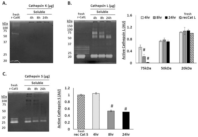
Figure 5. Cathepsins L and S remain active over longer periods of time in the presence of fibrin.
Zymography was used to determine the ability for fibrin to extend the activity time of cathepsins K, L, and S. (A) No active catK was detected in the supernatant after 4, 8, or 24 hours. (B) Bands of active catL appear with a cascading loss of higher molecular weight bands (100, 75, 50, and 37kDa) between 4 and 24 hours. (C) Active catS was only detected at 25kDa, its expected molecular weight, with the amount of active catS decreasing over 24 hours. (n=4, #p<0.0001)

