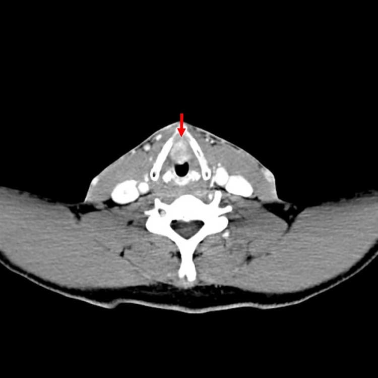
Fig. 1.

Axial CT of the neck demonstrates the 2.5-cm oval-enhancing mass along the true vocal cords with involvement of the anterior commissure, marked by arrow. The lesion does not demonstrate evidence of extralaryngeal spread, cartilage erosion, or paraglottic invasion. CT, computed tomography.
