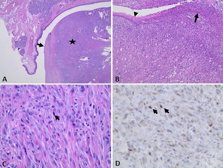
Fig. 5.
(A) Low power magnification shows a polypoid tumor (star) with ulcerated surface (arrow). (B) The tumor originates in the submucosa which is partially ulcerated (arrow head: overlying squamous epithelium; arrow: ulcerated mucosa). (C) The tumor is composed of highly malignant cells with marked atypia and pleomorphism. Mitoses (arrow) are readily identified. Note the absence of adipocytic differentiation. (D) MDM2 immunohistochemistry is positive (at various degrees) in the nucleus of tumor cells (arrows).

