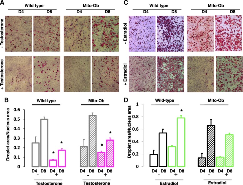
Fig. 9.
a, c Representative photomicrographs showing differentiation of subcutaneous preadipocytes from male and female Mito-Ob mice with and without respective sex steroid supplementation, as determined by Red Oil O staining (40×). b, d Respective histograms showing quantification of adipocyte differentiation. Preadipocytes from male and female wild-type mice were included as controls. Experiments were repeated for three to four times. *p < 0.05 represents significant differences between with and without sex steroid treatment within each experimental sub-groups (days, genotype and sex). D—days

