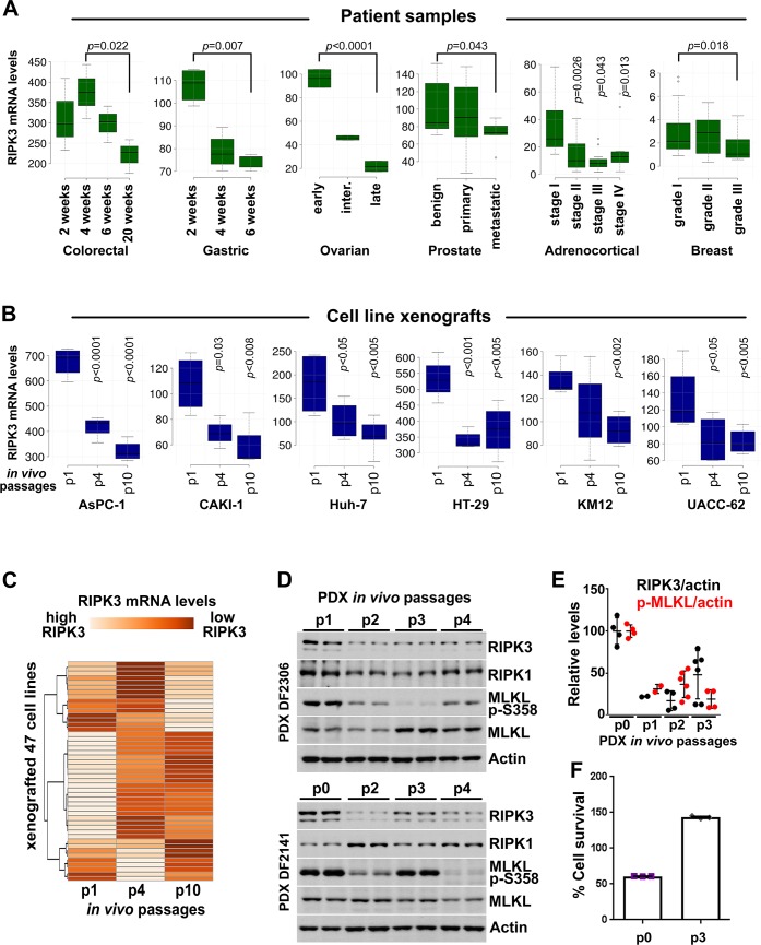
Fig 1. Necroptosis is induced in tumors in vivo and RIPK3 expression is progressively lost during tumorigenesis.
(A) RIPK3 mRNA levels are decreased during progressive stages of colorectal, gastric, ovarian, prostate, adrenocortical, and breast cancers. The results shown here are in part based upon data generated by the TCGA Research Network. See S6 Table for details about the studies. (B) RIPK3 expression is lost during progressive in vivo passages of mouse tumor xenografts of indicated cancer cell lines. (C) As in (B), except data from 47 different cell lines are presented as a heatmap. (D) Necroptosis is induced in tumors in vivo and RIPK3 expression is progressively lost during tumorigenesis. Necroptosis induction is determined by the MLKL p-S358. Ovarian PDX lysates obtained at the indicated in vivo passages were immunoblotted with the indicated antibodies. (E) Quantification of the RIPK3 and p-MLKL levels shown in (D) and their normalization to actin. (F) Ovarian PDX cells at indicated in vivo passages were cultured and treated with TSZ to induce necroptosis. Cell survival was determined 24 hours after treatment, using CellTiterGlo. The underlying data can be found in S1 Data. PDX, patient-derived xenograft; p-MLKL, phospho-MLKL S358; TSZ, TNFα+SM-164+zVAD.fmk.

