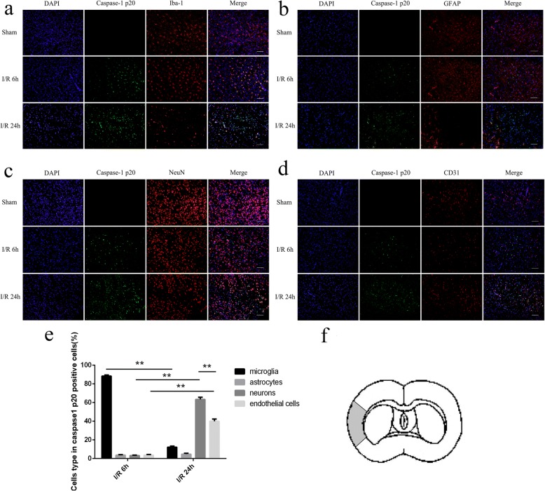
Fig. 1.
Cellular localization of cleaved caspase-1 after ischemia/reperfusion (I/R) injury. a–d The expression of caspase-1 p20 in microglia, astrocytes, neurons, and endothelial cells in sham rats and after cerebral I/R at 6 h and 24 h. e The percentage of different cell types in caspase-1 p20-positive cells 6 h and 24 h after cerebral I/R. f The ischemic area where the cleaved caspsae-1 was richly expressed. Bar = 100 μm. *p < 0.05, **p < 0.01

