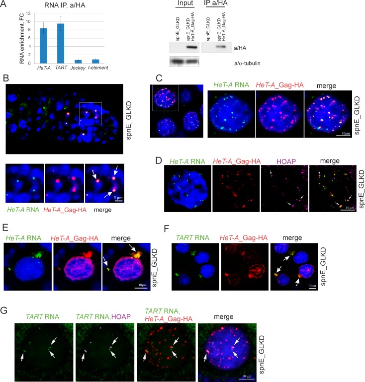
Fig 1. HeT-A RNA and HeT-A Gag protein form RNP in ovaries.
(A) RT-qPCR analysis of RNA immunoprecipitated with anti-HA from ovary lysates of nosGal4; UAS-HeT-A-HA; UAS-spnE_sh flies and control nosGal4; UAS-spnE_sh flies. Fold changes (FC) for RNA enrichments in RNA IP from ovaries of HeT-A-HA transgenic strain versus control are shown. rp49 was used for normalization. The error bars represent standard deviations (SD) of 2 biological replicas. Western blot analysis of immunoprecipitated HeT-A Gag-HA is shown to the right. The antibodies used for Western blotting are indicated to the right. (B-G) RNA FISH combined with immunostaining was performed on ovaries of nosGal4; UAS-HeT-A-HA; UAS-spnE_sh flies. (B) HeT-A RNA (green) colocalizes with HeT-A Gag-HA (red) in germarium. Enlarged region (white rectangle) is shown. HeT-A spheres (arrows) are observed in cystoblasts. (C) HeT-A RNA (green) colocalizes with HeT-A Gag-HA (red) in nurse cell nuclei. A fragment of a stage 7 egg chamber is shown to the left. An enlarged nucleus of a germline nurse cell (white rectangle) is shown. (D) HeT-A RNA FISH (green) combined with HeT-A Gag-HA (red) and HOAP (magenta) immunostaining in a nurse cell nucleus from a stage 7 egg chamber is shown. Telomeric localization of HeT-A RNP is shown by arrows. (E) HeT-A RNA (green) and HeT-A Gag-HA (red) form large aggregates (arrows) in the cytoplasm of nurse cells. A fragment of a stage 5 egg chamber is shown. For an entire egg chamber see Part A in S2 Fig. (F) TART transcripts (green) are co-localized with HeT-A Gag-HA (red) in the nurse cell cytoplasm (arrows). A fragment of a stage 7 egg chamber is shown. (G) TART RNA (green) colocalization with telomere-specific protein HOAP (magenta) is shown (arrows). HeT-A Gag-HA (red) partially colocalizes with TART RNA in nurse cell nuclei. Image of an individual nurse cell nucleus from a stage 6 egg chamber.

