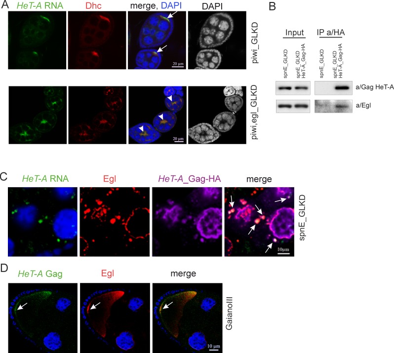
Fig 3. Egl participates in the transport of HeT-A RNPs in ovaries.
(A) Egl germline knockdown results in mis-localization of HeT-A transcripts. Egg chambers at stage 4–5 of oogenesis from piwi_GLKD (top panel) and double piwi-egl_GLKD (bottom panel) are stained with a/Dynein heavy chain (Dhc) (red). HeT-A RNA (green) is localized in the oocyte upon piwi_GLKD (arrows), but is mis-localized upon piwi,egl double GLKD (arrowheads). Ectopic Dhc is observed at this stage according to published data [40]. DAPI is shown separately in a grey. (B) Co-IP of HeT-A Gag. Western blot analysis of proteins immunoprecipitated with anti-HA from ovaries of spnE-GLKD flies (control) and nosGal4; UAS-HeT-A-HA; UAS-spnE_sh. Anti-HA immunoprecipitates HeT-A Gag-HA and Egl proteins. The antibodies used for Western blotting are indicated to the right and the antibodies used for co-IP are indicated above the IP lanes. (C) HeT-A RNA (green), HeT-A Gag-HA (magenta) and Egl (red) form aggregates (arrows) in the cytoplasm of the nurse cells in ovaries of nosGal4; UAS-HeT-A-HA; UAS-spnE_sh flies. The fragment of a stage 9 egg chamber is shown. For an entire egg chamber see Part A in S5 Fig. (D) HeT-A Gag (red) and Egl (green) co-localize at the posterior pole of the oocyte (arrows) at stage 10 in ovaries of GIII strain. A fragment of a stage 10 egg chamber including oocyte is shown. DNA is stained with DAPI (blue).

