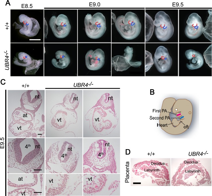
Fig 1. Mouse embryos lacking UBR4 die at midgestation associated with multiple developmental abnormalities.
(A) Gross morphology of +/+ and UBR4-/- mouse littermates at E8.5-E9.5. UBR4 mutants exhibit embryonic growth retardation and malformed pharyngeal arches. The red and blue lines represent the first PA and second PA, respectively (scale bar = 1 mm). (B) Anatomical illustration of the pharyngeal arch (PA) and heart of the mouse embryo at E9.0. First PA mesodermal cells (red) move to the developing heart tube for the right ventricular myocardium at E7.5–8.0, while second PA mesodermal cells (blue) migrate to the outflow tract (oft) of the myocardium at around E9.5-E10. (C) Hematoxylin and eosin (H&E) staining on cross sections of +/+ and UBR4-/- embryos at E9.5. nt, neural tube; at, atrium; vt, ventricles; 4th, fourth ventricle (scale bar = 100 μm). (D) H&E staining of the placenta of +/+ and UBR4-/- mouse littermates at E9.5 (scale bar = 500 μm).

