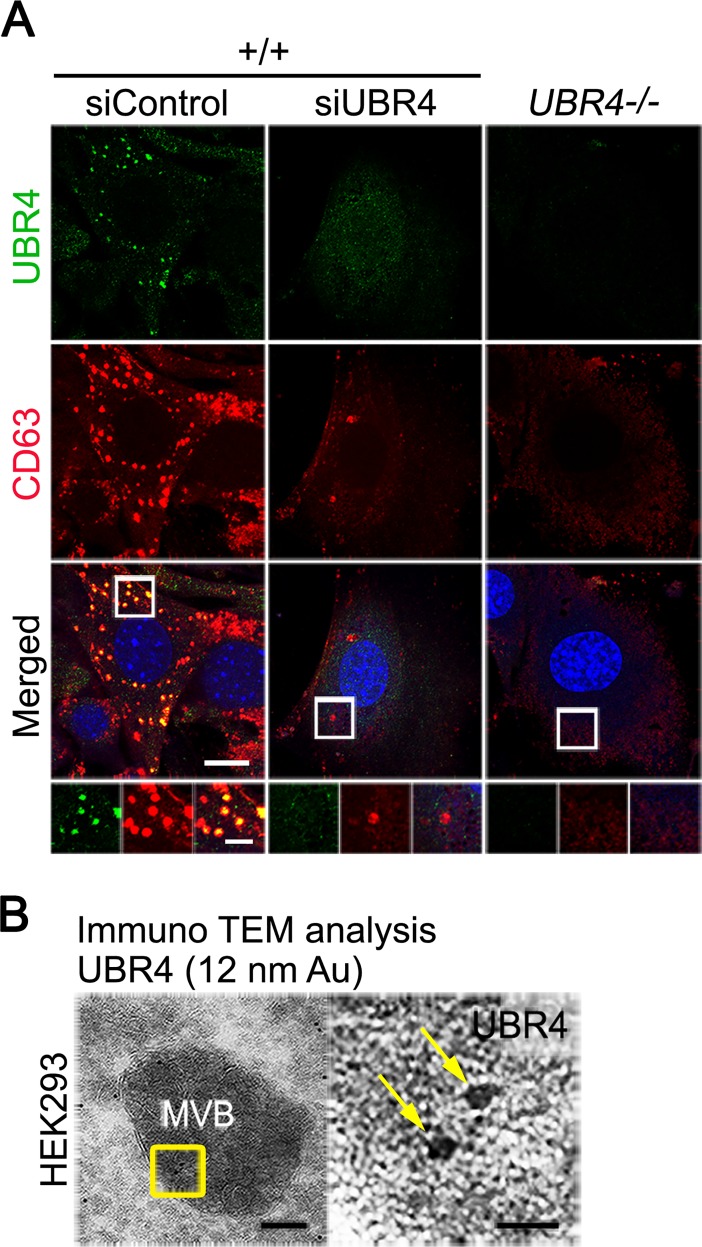
Fig 7. UBR4 is associated with MVBs and required for the formation of MVBs.
(A) Immunostaining of UBR4 and the MVB marker CD63 on siControl- and siUBR4-treated +/+ and UBR4−/− MEFs (scale bar = 15 μm and 300 nm, respectively). (B) Transmission electron microscopy (TEM) of HEK293 cells stably expressing UBR4-V5. Fixed cells were incubated with primary antibody against V5 followed by secondary IgG antibody labelled with 12 nm gold beads. Arrows indicate UBR4-V5 molecules located at an MVB (scale bar = 100 nm for left panel and 20 nm for right panel).

