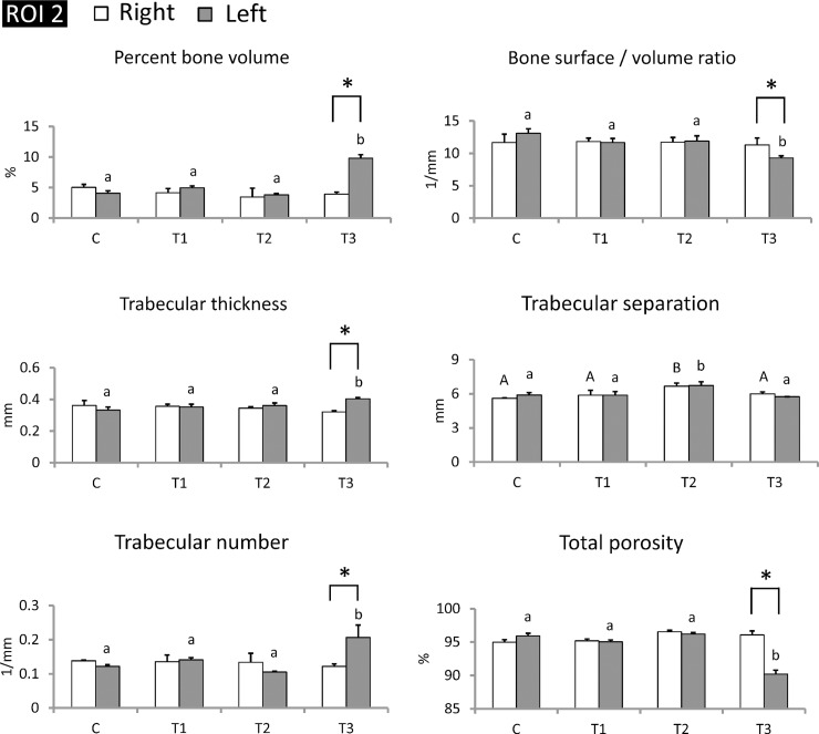
Fig 8. piPSC-derived osteoblast-like cells markedly improved trabecular microarchitecture in the left tibiae of T3 group at transplanted sites.
Transplanted cells ameliorated trabecular bone volume, thickness, separation, number, and total porosity at transplanted sites in the T3 group. The range of ROI (2) at transplanted sites. Left tibiae were subjected to cell transplantation, and right tibiae were maintained as internal control. C, control; T1, treatment 1; T2, treatment 2; T3, treatment 3. AB/ab: Values in the same site with different letters indicate significant differences. *: P < 0.05 versus Right (Duncan’s multiple range test). N = 3 in each group.

