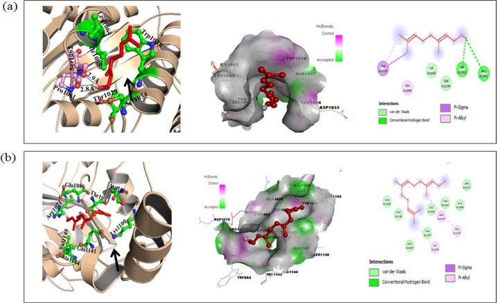
Fig 5. Molecular docking of Ger with CaCdr1p.
(a) Cartoon model of Cdr1p protein with Ger (left panel), surface view of CaCdr1p with Ger (middle panel) and 2D schematic diagram showing interactions of Ger to the CaCdr1p. Residues involved in hydrogen bonding, Vander Waals interactions, Pi-sigma and Pi-alkyl are represented in different color indicated in inset (right panel). (b) Cartoon model of CaCdr1p with Farnesol (known inhibitor) (left panel), surface view of CaCdr1p with Farnesol (middle panel) and 2D schematic diagram showing interactions of Farnesol to the CaCdr1p. Residues involved in hydrogen bonding, Vander Waals interactions, Pi-sigma and Pi-alkyl are represented in different color indicated in inset (right panel).

