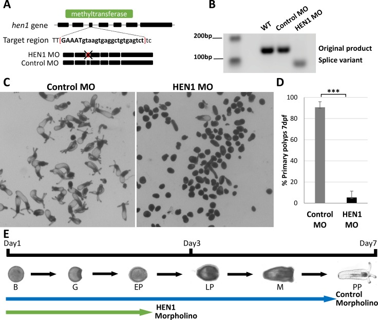
Fig 3. HEN1 is essential for Nematostella development.
(A-B) Schematic diagram of MO targeting region on the hen1 gene of Nematostella. The MO is designed to target hen1 exon-intron junction located in methyltransferase domain (green). This MO impaired the splicing by deleting 3rd exon of hen1. The splicing variation was validated by PCR. Due to deletion of 3rd exon, the band in HEN1 MO-injected embryos shifted down. In contrast, the bands in control MO-injected embryos and wildtype presented the expected size. (C-D) Animals injected with control MO developed to primary polyps after 7 dpf. In contrast, animals injected with HEN1 MO stopped developing prior to metamorphosis (D) ~90% of HEN1 depleted animals did not reach primary polyp stage at 7 dpf, triplicates, n = 300, ***P < 0.001 (Student’s t-test). (E) The timeline of Nematostella development and the relative progress of control and HEN1 MO-injected animals. B = Blastula; G = Gastrula; EP = Early Planula; LP = Late Planula; M = Metamorphosis; PP = Primary Polyp.

