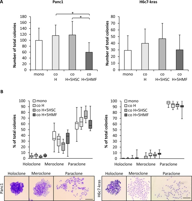
Figure 1. The hepatic microenvironment supports self-renewal of PDECs.
Panc1 and H6c7-kras cells were either monocultured (mono) or indirectly cocultured in different experimental hepatic environments, consisting of hepatocytes alone (co H) or hepatocytes enriched with 5% HSC (co H+5HSC) or 5% HMF (co H+5HMF), respectively, for 6 days. (A, B) After 6 day culture under the described conditions, PDECs were detached and 400 cells seeded for colony formation which was assessed after crystal violet staining on day 10. Only colonies containing more than 50 cells were counted and (A) the total number of colonies and (B) the proportion of different colony types of total number of colonies were determined. Due to reasons of clarity, significant differences are not marked in this chart. Data are presented as mean and standard deviation or median and quartiles (Q1 as 25% and Q3 as 75%) of 6 to 7 independent experiments. Below, representative images of crystal violet-stained holo-, mero- and paraclones of Panc1 and H6c7-kras cells are shown. Scale bar 250 μm. * indicates statistically significant differences (p ≤ 0.05).

