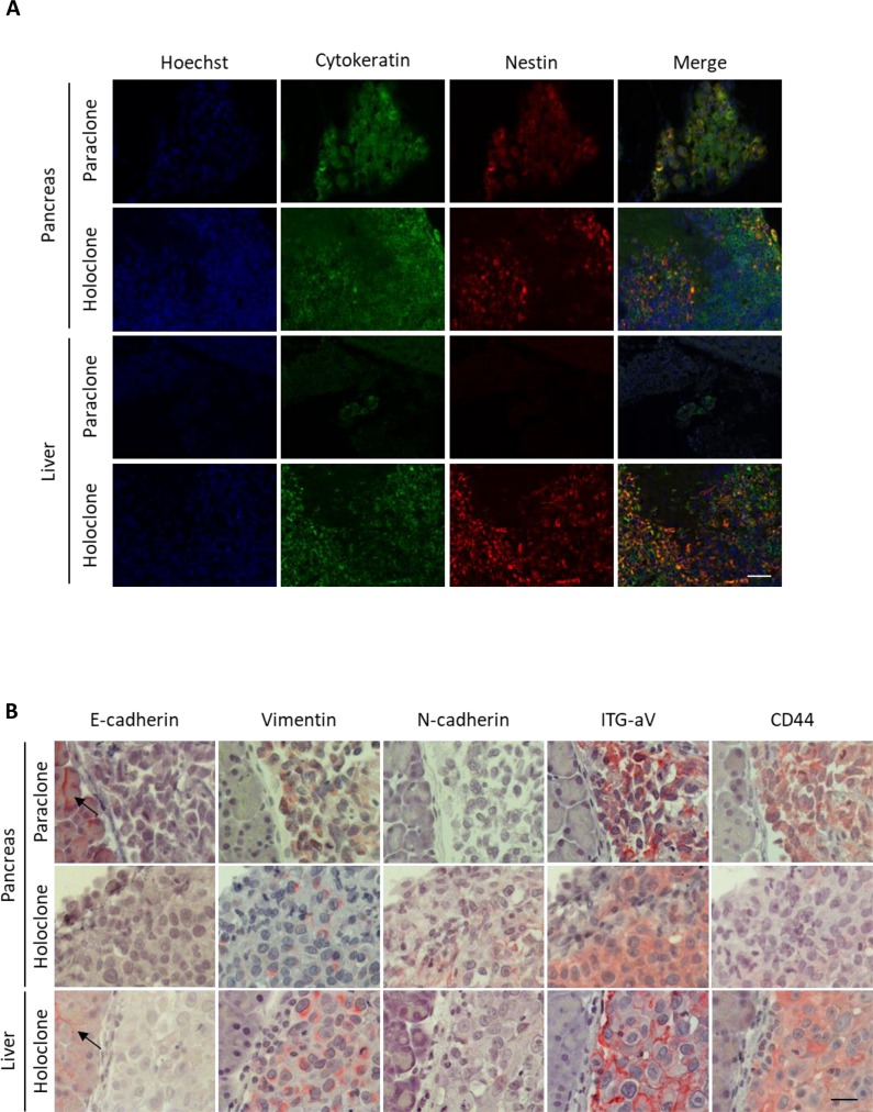
Figure 4. Panc1 holoclone tumors exhibit a pronounced mesenchymal phenotype along with elevated Nestin expression.
Tumorigenic potential of Panc1 holoclone and paraclone cells derived from an HSC-enriched coculture was investigated by intrasplenic inoculation of 104 holoclone or paraclone cells into SCID-beige mice (n = 10 animals/cell type). Formalin-fixed paraffin-embedded tissue sections of pancreatic and corresponding liver sections were (A) immunofluorescently co-stained for human Cytokeratin (green) for detection of human Panc1 cells and the CSC-marker Nestin (red). Hoechst staining was performed to mark nuclei (blue). Scale bar 50 μm. (B) Pancreatic and hepatic tissues were immunohistochemically stained for E-cadherin, Vimentin, N-cadherin, Integrin alpha V (ITG-aV) and CD44. Arrows indicate E-cadherin-expressing non-neoplastic tissue cells. Haemalumn staining was performed to counterstain nuclei. Scale bar 25 μm.

