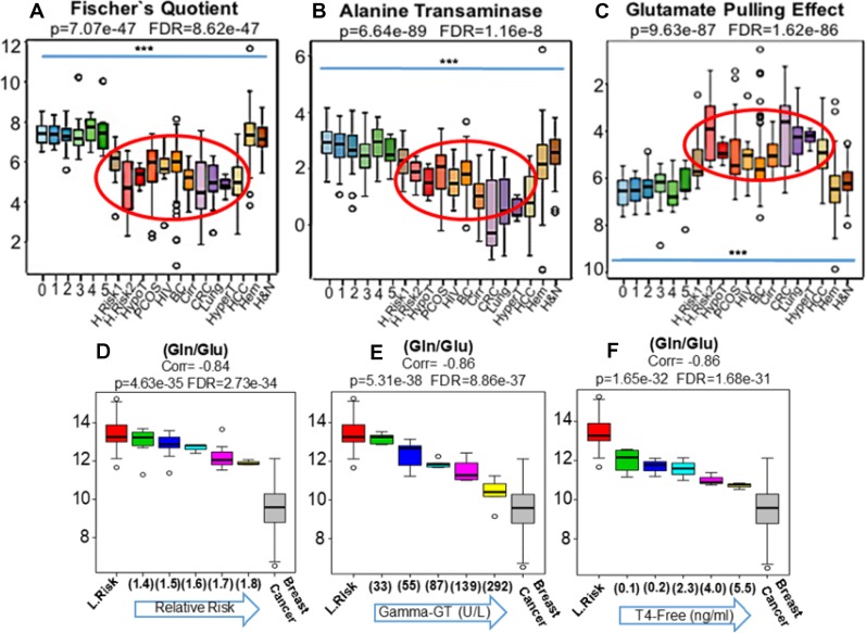
Figure 3. The MYC-coordinated and malignancy-associated increase in glutamate production, after in vitro shortages of glucose, was previously described as the “glutamate pulling effect” [11].
The ratio (Glu/Hexoses) was adopted here as a proxy for this metabolic shift that, in fact, was clearly replicated in the blood (Red eclipse) of patients harboring adenocarcinomas (BC, CRC, Lung and HCC) as well as in women at higher risks of breast cancer development (R.R. = 1.5 H.Risk 1) and (R.R. = 1.8 H.Risk 2) and in individuals with PCOS (C). Of note, neither in the population-based controls depicting progressive metabolic syndrome (0 to 5) or in patients harboring non-glandular tumors such as leukemias, myelomas and lymphomas (Hem) as well H&N squamous cells carcinomas revealed significant changes in the ratio (Glu/Hexoses) (C). Since the results generated by the Fischer's quotient (A) were persistently suggesting liver dysfunctions in patients harboring glandular malignancies, we also compared our findings to well established conditions of liver dysfunctions such as cancer-free patients with HCV-induced cirrhosis (Cirr), patients with hypo (HypoT) and hyperthyroidism (HyperT), as thyroid dysfunction is very frequently associated with liver metabolic abnormalities as well as to increased risks of breast cancer [23–25]. Similarly, we also analyzed HIV patients due to increased risks of cancer development and because of the direct HIV influence on liver function (26). Results revealed concordance between the blood phenotypic profiles of cancer-free patients with cirrhosis, thyroid dysfunction and HIV infection with study participants at elevated relative risks of cancer, those with polycystic ovary syndrome (PCOS) and patients harboring glandular malignancies (A–C Red ellipses). To explore in more details the relations among malignancy, thyroid and liver function, we further divided our cancer-free groups according to: i- increasing relative risks of breast cancer (from 1.4 to 1.8) (D), ii- rising levels of gamma-GT (from 33 to 392 U/L) (E) and iii- cumulative values of free-thyroxin (from 0.1 to 5.5 ng/mL) (F) and compared the findings to women at lower risks of breast cancer (L.Risk) as well as participants with stage III invasive disease (Breast Cancer). Results revealed that the same pattern generated by the ratio (Gln/Glu) when applied to cancer-free high risk participants (D), could be precisely recapitulated in blood of cancer-free women according progressive values of gamma-GT (E) and free-T4 (F). *** Indicates p < 0.001 (H&N: Head and Neck Cancer).

