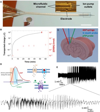
Fig. 1. Overview of the μFIP probe.

(A) Implanted end of the device (inset scale bar, 100 μm; outside scale bar, 1 mm). (B) Net transported charge across the ion bridge when actively pumping GABA at 1 V (line, left axis), [GABA] passively diffused out of the device when no voltage was applied (open symbols, right axis), and [GABA] actively pumped out of the device at 1 V (closed symbols, right axis). (C) Schematic showing placement of syringe for 4AP injection, Si depth probe, and the μFIP probe in the hippocampus. (D) Conceptual illustration showing a proposed effect of 4AP on K+ channels and action potentials (31) along with the analogous effects of GABA. (E) Representative recording of intense SLEs following injection of 4AP at two different time scales.
