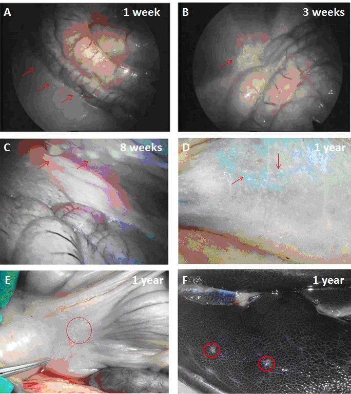
Figure 2. Graft retrieval from the peritoneal cavity of pigs.

Laparoscopy images show the presence of large number of free transparent microcapsules at week 1 (A) and week 3 (B) respectively. At week 8 the number of free microcapsules was less and most capsules were found adhering to abdominal structures (C). No free microcapsules were seen by 1 year post-transplantation and papules, consistent with clusters of adherent microcapsules, were visible at a number of sites including posterior peritoneal wall (D), omentum (E) and liver (F).
