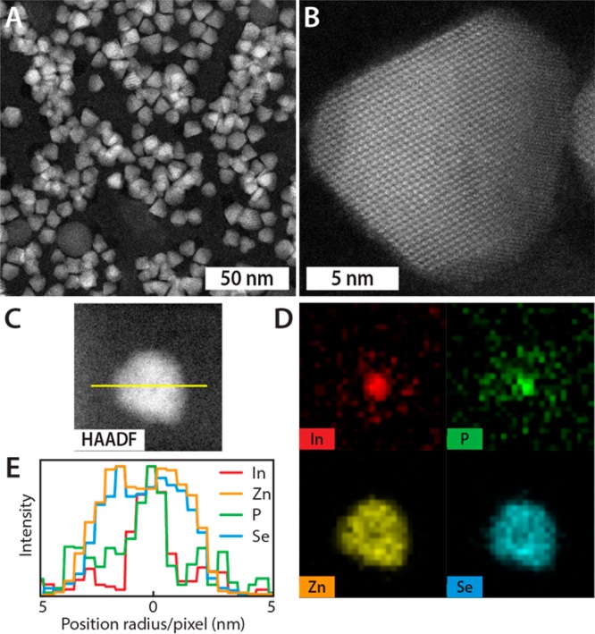
Figure 1.

Chemical and structural characterization of InP/ZnSe core/shell QDs. (a and b) HAADF-STEM images of InP/ZnSe core/shell QDs, with an average InP core diameter of 2.9 nm and a total core/shell diameter of 9.6 ± 1.1 nm (mean ± standard deviation), at (a) low and (b) high magnification. The high magnification depicts the ZnSe crystal along the [110] direction. (c and d) HAADF-STEM image of a single InP/ZnSe QD and corresponding EDX maps providing evidence for an InP core diameter of approximately 3 nm and a ZnSe shell of 3–4 nm. (e) Elemental line scan along the yellow line through the InP/ZnSe QD shown in panel c. The core/shell structure of the QD is clearly resolved, since In and P are present primarily in the center of the nanocrystal, while Zn and Se are distributed throughout the QD. The diffuse and weaker background signal of P is ascribed to trioctylphosphine (TOP), which acts as a ligand.
