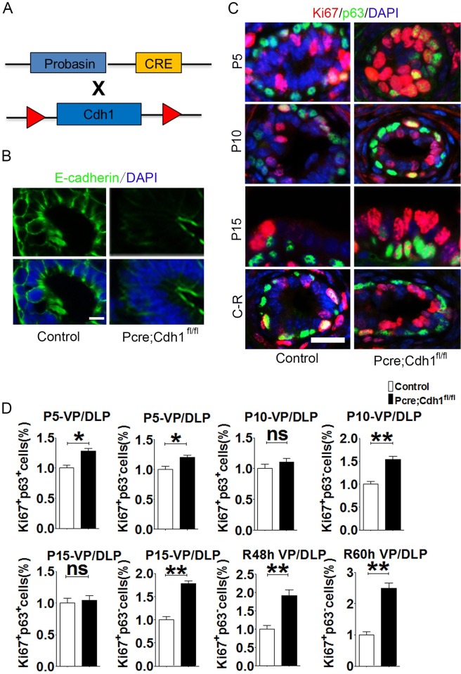
Fig 1. E-cadherin ablation leads to a hyperproliferative phenotype of prostatic luminal cells.
(A) The schematic illustrates the generation of a mouse model with prostatic specific knockout of E-cadherin. (B) The efficiency of conditional E-cadherin deletion is confirmed by immunofluorescent staining. (C) Analysis of cell proliferation in developing prostates (P5, P10 and P15) or regenerating prostates (48h and 60h after androgen replacement) by immunofluorescent co-staining of proliferative marker Ki67 and basal cells marker p63 (Scale bars are 20μm for (B) and (C)). (D) Quantification of mitotic epithelial cells from ventral and dorsolateral prostatic lobes at different development stages or regeneration time points (n = 3, Student’s t-test, ***P<0.001,**P<0.01, *P<0.05,error bars = SEM.).

