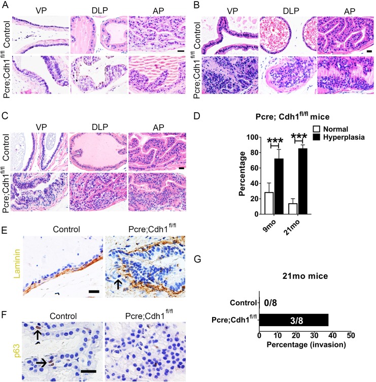
Fig 2. E-cadherin knockout results in development of invasive prostate adenocarcinoma.
(A) H&E staining shows that multilayered epithelia structure and hyperplastic lesions can be found in 4-month old E-cadherin knockout mouse prostates. (n = 4) (B) Nine-month old E-cadherin knockout mouse prostates develop high-grade murine intraepithelial neoplasia. (control n = 5, Pcre;Cdh1fl/fl n = 6) (C) H&E staining shows adenocarcinoma in the prostates of 21-month-old Pcre;Cdh1fl/fl mice. (n = 8) (D) Quantification of hyperplastic lumens in total prostate lumens from 9- or 21-month-old mice. (E) Immunohistochemistry staining of laminin indicates the disruption of the basement membrane and invasion of epithelial cells into the surrounding prostatic stroma in adenocarcinoma from Pcre;Cdh1fl/fl prostates. (F) Loss of p63+ basal cells in adenocarcinoma from Pcre;Cdh1fl/fl prostates (Scale bars are 20μm). (G) Quantification of the invasive incidence from 21-month-old mice. (Student’s t-test, **P<0.01, *P<0.05, error bars = SEM. 9 month, control n = 5, Pcre;Cdh1fl/fl n = 6; 21month, n = 8).

