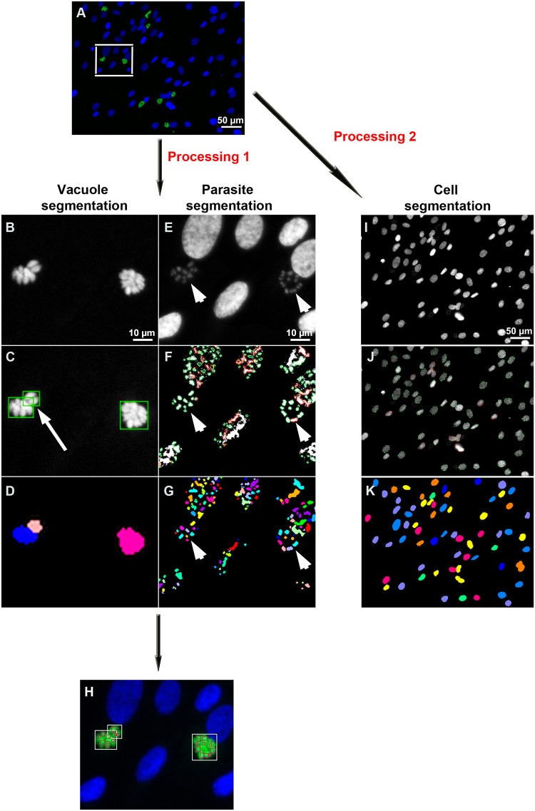
Fig 1. Images processing steps.
A: Example of raw image acquired with the automated ScanR Olympus software. B-H: Cropped images to explain the image process analysis. B-D: Image processing to detect Toxoplasma parasitophorous vacuoles using the YFP2 /Alexa-488 channel. E-G: Image processing to detect parasites using the Hoechst-33342 channel. C, F: Images were filtered, the background was subtracted and a watershed algorithm was applied to identify both the vacuoles and the parasites. D, G: A binary vacuole mask and a parasite mask were generated. H: Masks were merged to identify parasites inside their vacuoles. I-K: Host cell nuclei processing. I: Host cells were detected using the Hoechst-33342 channel. J: Images were filtered, the background was subtracted and an edge algorithm was used to segment host cells. K: A binary host cell mask was generated.

