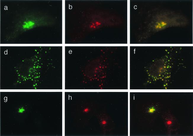
Figure 3.
Colocalization of TGN38-pHluorin GFP with TGN markers. Bronchial epithelial cells were cotransfected with TGN38-pHluorin GFP and myc-tagged α2,6-sialyltransferase Sttyr. (a–c) IB3–1 cell. (a) GFP fluorescence. (b) Immunofluorescent visualization of myc-tagged α2,6-sialyltransferase Sttyr by using anti-myc Ab and secondary Alexa 568-conjugated Ab (red fluorescence). (c) Merged images a and b. (d–f) IB3–1 cell treated as in a–c with the addition of 20 μg/ml of nocodazole for 1 h. (d) GFP fluorescence as in a. (e) Myc-tagged α2,6-sialyltransferase Sttyr (red fluorescence). (f) Merged images d and e. (g–i) TGN-38-pHluorin GFP colocalization with human syntaxin-6 in IB3–1 cell. (g) GFP fluorescence. (h) Immunofluorescent visualization of anti-human syntaxin-6 (primary Ab) by using secondary Alexa 568-conjugated Ab (red fluorescence). (i) Merged images g and h. Appearance of C38 and S9 cell was identical to IB3–1 cells.

