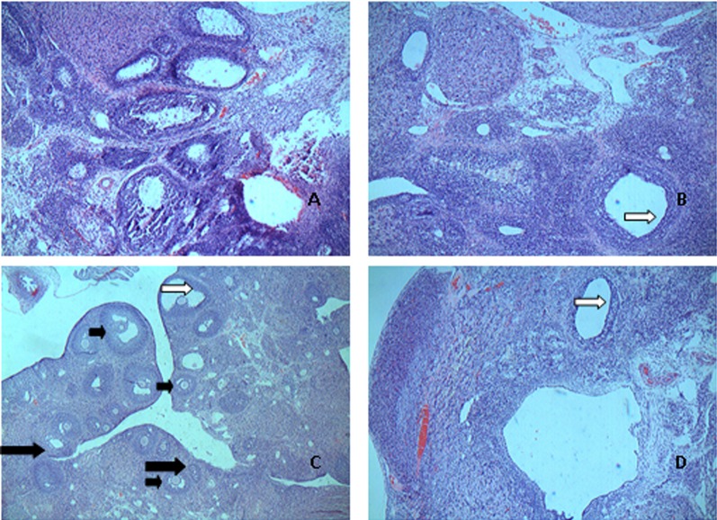
Fig. 1.
Sections of ovaries from control group, model group, high dose group and estrogen group stained with hematoxylin and eosin (Original magnifieation: × 100). Different stages are indicated by different arrows (small black arrows, mature follicle; white arrows, ovarian theca cell; and large black arrows, corpus luteum). A: CG; B: MG; C: H-Gen; D: EG.

