Figure 5.

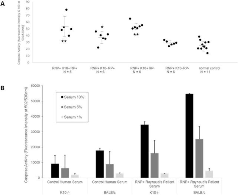
K10-dependent endothelial apoptosis. A. HUVEC were exposed for 24 hours to 10% dilutions of sera from healthy subjects (Ctrl) or RNP+ patients with or without Raynaud’s (RP+ or -) and anti-K10 IgG antibodies (ELISA > 2 S.D. above healthy subject mean to be anti-K10+), loaded with CellEvent Caspase 3/7 Green Detection Reagent (10μM/ml) and assayed for green absorbance in 24 hours in duplicate wells. Anti-K10+ sera induced high levels of caspase activity (an indicator of apoptosis) from patients with or without RP. ** t test p < 0.041 versus K10-RP+ sera, p < 0.003 versus K10-RP- sera, and p <= 0.0001 versus Ctrl K10- sera; * t test p < 0.048 versus K10-RP- sera and p < 0.009 versus Ctrl K10- sera. Results were representative of 3 separate experiments. B. Endothelial cell cultures concurrently generated from BALB/c, and K10-knockout BALB/c mice (see Methods) were exposed to dilutions of K10- control serum or K10+ RNP+ RP+ serum for 24 hours, and assayed for caspase activity as above (representative of 2 separate experiments). The Raynaud’s serum induced increased dose-dependent caspase activity in both K10-intact and K10−/− cells compared to control serum, but much more so in K10-intact cells.
