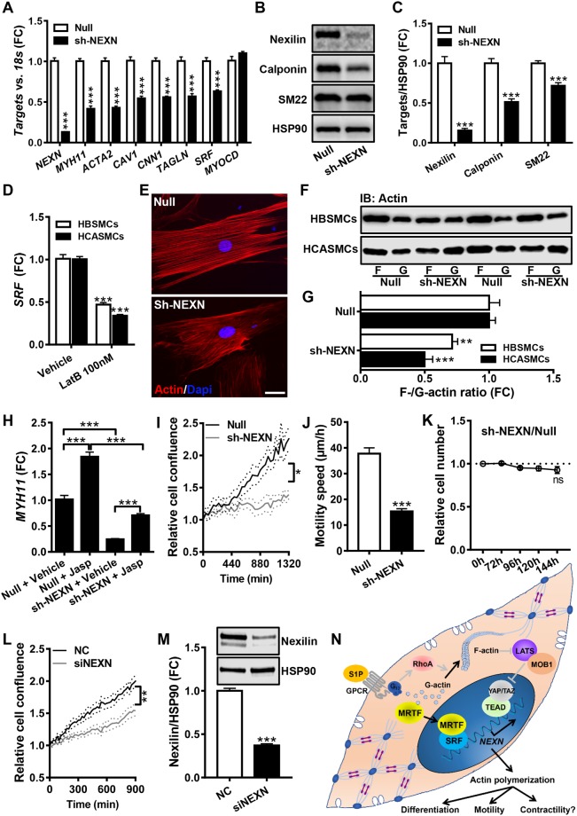
Figure 7.
Knockdown of Nexilin/NEXN reduces actin polymerization and SMC marker expression. NEXN was silenced using a short hairpin adenoviral construct (sh-NEXN) and the contents of NEXN and a number of SMC differentiation markers were examined at the mRNA (panel A, N = 6–9) and protein levels (panels B and C, N = 12). Panel D shows that depolymerization of actin using LatB reduces SRF expression in both human bladder and coronary artery SMCs (HBSMCs, N = 8; HCASMCs, N = 9). Panel E shows phalloidin staining of actin filaments in control cell (Null) and after NEXN silencing (sh-NEXN). The scale bar represents 50 μm. Panel F shows a sedimentation assay to determine filamentous (F-) and globular (G-) actin in control cells (null) and after silencing of NEXN. Panel G shows the normalized F- to G-actin ratio in bladder and coronary artery SMCs (HBSMCs, N = 12; HCASMCs, N = 6). Panel H shows that polymerization of actin using jasplakinolide (Jasp) increases MYH11 in HBSMCs in control conditions and after NEXN silencing (N = 6). Panel I shows cell density at different times following the creation of a cell-free area in the culture dish (N = 9–10). Panel J shows the speed of cell movement within the cell-free area (N = 19 and 22 motile cells in Null and sh-NEXN, respectively). Panel K shows a cell viability assay comparing control and NEXN-silenced cells (N = 12 throughout). The effect of NEXN silencing on cell migration was confirmed using an independent siRNA in L (N = 9). The associated repression of the NEXN protein is shown in (M) (N = 6). The schematic illustration in panel N summarizes our findings regarding the transcriptional control of NEXN and its impact on actin polymerization and cell motility. Several aspects of this model, including the involvement of a (G) protein-coupled receptor in the S1P effect, were not directly tested here, and they are thus drawn in grey.

