Fig. 5.
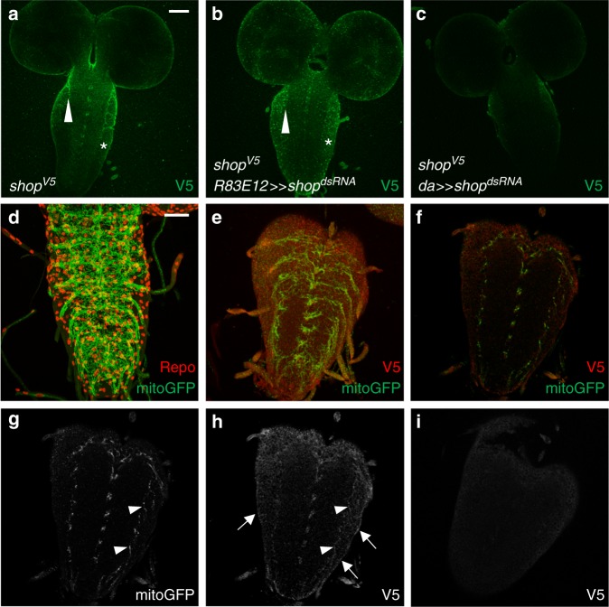
Shopper localizes to mitochondria of the ensheathing glia. a ShopperV5 expression (green) in a third-instar larval brain. Note the enrichment of Shopper expression in the ensheathing glia (arrowhead) and the weak expression in the CNS cortex (asterisk). b Upon expression of shopperdsRNA in all ensheathing glial cells, Shopper expression cannot be detected in the ensheathing glia anymore (arrowhead), but the expression in the cortex glia remains (asterisk). c Upon ubiquitous expression of shopperdsRNA using the da-Gal4 driver Shopper expression is abolished. d The organization of mitochondria in ensheathing glial cells in third-instar larval brains as revealed by expression of UAS-mitoGFP using R83E12-Gal4 (green). Repo staining (glial nuclei, red) and HRP staining (neuronal membranes, blue) are shown as well. Note that this is a projection of a confocal stack of a paraformaldehyde (PFA)-fixed specimen. e Endogenous Shopper expression as revealed by expression of an V5-tagged Shopper protein (red) in an animal expressing UAS-mitoGFP in the R83E12-Gal4 pattern (green). Note that detection of V5 only works after fixation with Bouin’s fixative. f–h Single focal planes of the same stack as in (e); staining as indicated. Enrichment of Shopper expression can be detected in neuropil-associated glial cells (arrowheads) and the surface glial cells (arrows). i Control staining of a wild-type nervous system for V5 expression following Bouin’s fixation. Note the high background in the CNS cortex. Scale bars are 20 µm

