Fig. 5.
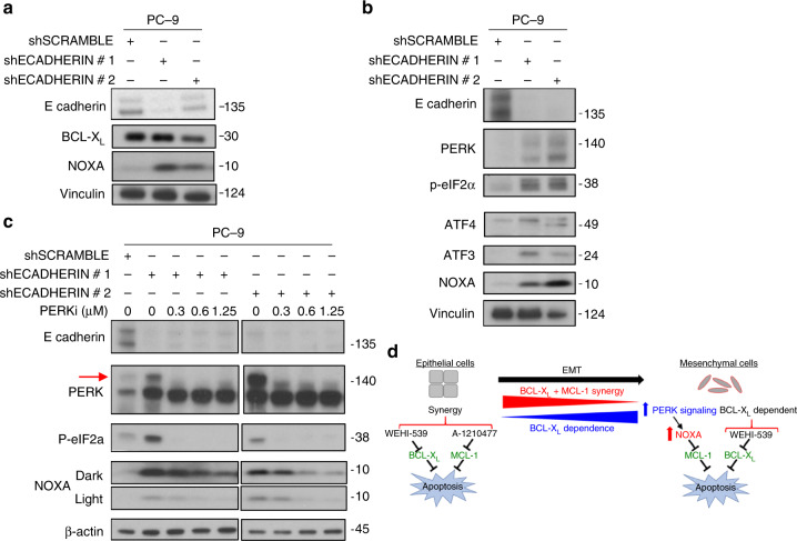
EMT induces NOXA expression and BCL-XL dependence via the PERK pathway. a Western blot data of PC-9 cells transduced with shECADERIN to induce an EMT. Expression levels of E-CADERIN, BCL-XL, NOXA, and Vinculin are compared via western blotting in cells after a 3-day selection in puromycin. b Expression levels of PERK signaling markers in cells forced to undergo EMT. PC-9 cells were transduced with shECADERIN and after 3 days in puromycin, markers of the PERK signaling pathway (PERK, p-eIF2α, ATF4, ATF3, NOXA) were compared using western blotting. c Blocking PERK signaling prevents EMT-induced NOXA expression. PC-9 cells were transduced with shECADHERIN or control and immediately incubated in the indicated concentrations of the PERK inhibitor (PERKi) GSK2606414 for the remainder of the 3 days in puromycin. Loss of the PERK autophosphorylation band (red arrow) indicates that PERKi is effective at the concentrations used. d Model: Epithelial cells tend to exhibit a synergistic co-dependence on BCL-XL+MCL-1, and therefore respond best to a combination of WEHI-539 and A-1210477. As cells become more mesenchymal like, either initially or via forcing an EMT, there is an increase in PERK signaling and PERK-dependent NOXA expression. This higher expression of NOXA renders cells more dependent on BCL-XL alone because the relatively higher amounts of NOXA neutralize MCL-1 in these cells. As such, the more mesenchymal cells are more responsive to WEHI-539 used as a single agent

