Figure 7.
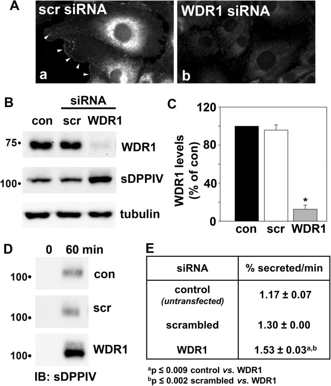
WDR1 knockdown enhances secretion. (A) Clone 9 cells were transfected with scrambled (scr) (panel a) or WDR1-specific siRNA duplexes (panel b), incubated for 48 h and immunolabeled for WDR1. Arrowheads are marking WDR1-positive cell surface patches in panel a. Note the considerable decrease in labeling in panel b. (B) Clone 9 cells were transfected with scrambled (scr) or WDR1-specific siRNA duplexes and incubated for 24 h. The control and transfected cells were infected with recombinant adenoviruses encoding V5-tagged sDPPIV and incubated an additional 24 h. Total cell lysates were immunoblotted for WDR1, sDPPIV and tubulin as indicated. Molecular weight standards are indicated on the left (in kDa). (C) The relative levels of WDR1 were calculated from densitometric analysis of immunoreactive bands on blots as shown in (B). Control values were set to 100%. Values are expressed as the mean ± SEM from three independent experiments. *p ≤ 0.001. (D) sDPPIV-expressing cells were rinsed five times with prewarmed serum-free medium and then reincubated in serum-free medium. At 0 and 60 min after reincubation, aliquots of media were collected and analyzed for sDPPIV secretion by immunoblotting with anti-V5 antibodies. (E) The percent sDPPIV secreted relative to the total expressed (B) was calculated from densitometric analysis of immunoreactive bands on blots as shown in (D) and the rate calculated. Values are expressed as the mean ± SEM from at least three independent experiments.

